Hospitals
Medical FacilitiesThe hospital has introduced a variety of state-of-the-art equipment for early detection and treatment in the hospital.
endoscope
In recent years, the number of patients with colorectal cancer has increased in Japan, and the mortality rate of colorectal cancer is now the highest among women. On the other hand, thanks to advances in medicine, colorectal cancer can be almost completely cured if detected at an early stage. Our clinic uses a thin and soft camera, which allows patients to undergo examinations with less burden.
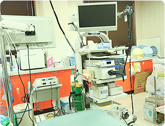
Features of our endoscope
Less abdominal tightness and pain
Carbon dioxide air delivery is introduced to reduce abdominal tightness and pain.
Free choice of nasal or oral
You can choose either nasal or oral insertion, whichever you prefer. For those with a strong gag reflex, nasal insertion is recommended.
Can be inspected while sleeping
The examination can be performed while the patient is sleeping with an intravenous drip to alleviate pain.
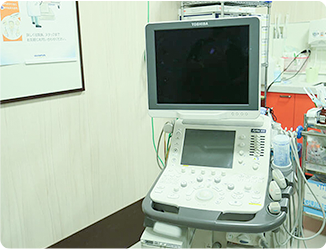
echo
Echocardiography is an examination in which ultrasound is applied to the affected area and the echoes are visualized. It is suitable for examining the stomach, intestines, liver, gallbladder, pancreas, kidneys, spleen, and great vessels. It is particularly useful for detecting gallstones and early-stage liver cancer.
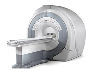
MRI
MRI (Magnetic Resonance Ima gi ng) is an examination that uses a strong magnetic field and weak radio waves (radio waves) to obtain a tomographic image of the human body.
Although similar in appearance to CT, MRI does not expose the patient to radiation and provides better contrast of human tissues than CT, especially for lesions of the brain, spine, articular cartilage, fat, and abdominal and pelvic organs.
Depending on the body part, blood vessels can also be visualized without the use of contrast media. In June 2023, we upgraded our MRI system to a GE SIGNA Explorer Newgrade l. ST. Compared to the previous system, the time required for examination has been shortened and clearer images can be obtained with the latest system, which also reduces the burden on patients during examination.
Bone densitometry (DXA)
Bone densitometry is a test to measure bone mineral density (bone mineral content) using X-rays. It can be used to diagnose osteoporosis, etc., and is also used for follow-up observation and to determine the effectiveness of treatment.
We use the highly accurate DXA (Dual Energy X-ray Absorpti ometry) method for our examinations.
The basic measurement sites are the lumbar spine and femoral neck, where the risk of fracture is particularly high. The measurement results are compared with those of young adults of the same age group. Comparison with the Young Adult Mean (YAM) is used to diagnose osteoporosis. A bone mass loss of 70~80% is diagnosed as osteoporosis, while a bone mass loss of less than 70% is diagnosed as osteoporosis.
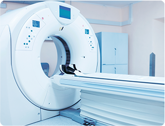
CT (computerized tomography)
CT stands for Computed Tomography, an examination that uses X-rays to obtain a cross-sectional image of the human body.
The hospital is equipped with a multi-slice CT (80 rows) and can quickly respond to urgent examinations. We also perform a variety of examinations ranging from physical checkups to whole body examinations. It is possible to take very thin tomographic images, enabling detailed observation of the inside of the body from various directions.
The workstation can also be used to create 3D images of bones and blood vessels, which facilitates three-dimensional observation and is helpful in explaining the results to patients. There are two types of examinations: simple CT examinations that do not use contrast media, and contrast CT examinations that use contrast media. Contrast is used to make organs and blood vessels more clearly visible, and is used only when it is deemed necessary for diagnosis.